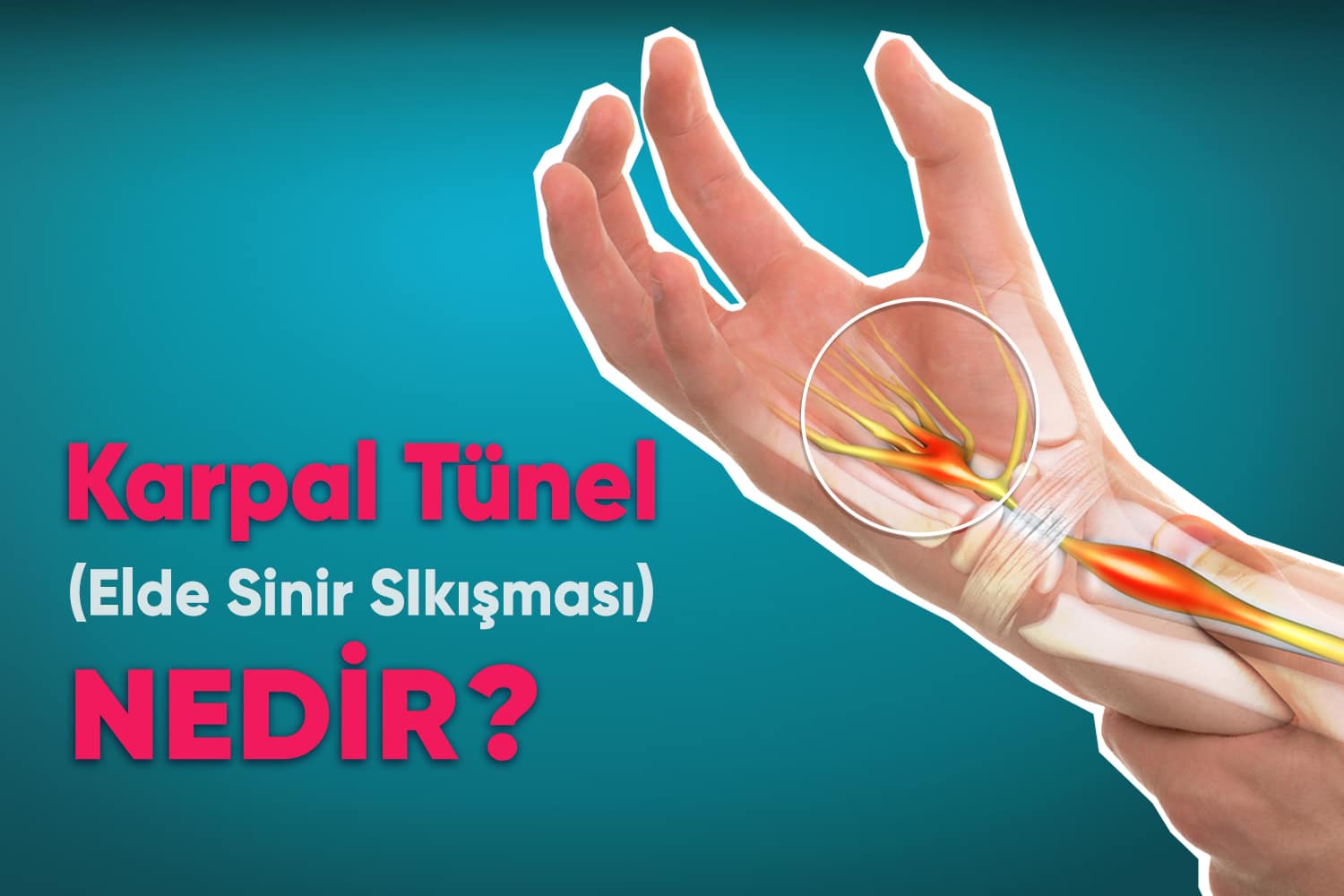
The carpal tunnel is a rigid fibro-osseous canal at the base of the wrist. Carpal Tunnel Syndrome arises when elevated intracanal pressure compresses the median nerve, compromising motor and sensory innervation to the hand.
Carpal tunnel syndrome characteristically presents with progressive neuropathic symptoms:
Numbness & Tingling: Patients experience burning and paresthesias in the thumb, index, middle, and ring fingers, frequently waking them at night or during prolonged wrist flexion.
Hand Weakness: Chronic motor fiber impairment causes grip weakness, thenar muscle atrophy, and frequent dropping of household objects.
Any process that reduces tunnel volume or increases tendon bulk—such as distal radius fractures, arthritis, or flexor tenosynovitis—compresses the median nerve.
Multiple anatomical, endocrine, and ergonomic risk factors are strongly linked to median nerve compression:
Bunlar:
1. Anatomical Biomechanics: Wrist fractures or small carpal canal dimensions increase mechanical pressure on the nerve.
2. Biological Sex: CTS occurs up to three times more frequently in females due to smaller baseline canal dimensions.
3. Neuropathic Illness: Chronic metabolic conditions such as diabetes mellitus heighten neural susceptibility to compression.
4. Inflammatory Disorders: Rheumatoid arthritis and tenosynovitis cause hypertrophic tissue swelling.
5. Pharmacological Associations: Certain medications, including aromatase inhibitors used in oncological therapies, are linked to increased incidence.
6. Fluid Retention: Elevated BMI, pregnancy, and menopause cause intracanal fluid retention.
Accurate diagnosis combines clinical provocative tests with electrodiagnostic evaluations:
Symptom Profile: Reviewing anatomical sensory loss and nocturnal flare-ups.
Physical Provocative Tests: Phalen’s test, Tinel’s sign, and Durkan’s compression test elicit localized neuropathic responses.
Radiographs: Wrist X-rays rule out osteoarthritic spurs or carpal fractures.
Neuromuscular Ultrasound: Accurately measures median nerve swelling and cross-sectional area.
Electromyography (EMG): Evaluates electrical motor unit activity to grade thenar denervation.
Nerve Conduction Velocity (NCV): Quantifies sensory latency and conduction speed across the carpal ligament.
Treatment Options
Timely intervention prevents irreversible nerve damage. Non-operative options include neutral splinting, anti-inflammatory therapies, and ergonomic pacing:
Taking frequent ergonomic rest intervals during typing,
Avoiding sustained forceful wrist flexion and heavy vibration,
Applying targeted cryotherapy to reduce localized soft-tissue swelling.
Comprehensive conservative therapies include neutral wrist immobilization splints, targeted pharmacotherapy, and ergonomic task pacing. When mild-to-moderate symptoms have been present for less than ten months, non-operative care yields substantial clinical improvement. If progressive numbness or manual clumsiness occurs, specialized orthopedic evaluation is strongly recommended.
NON-SURGICAL CONSERVATIVE THERAPIES:
Non-surgical therapies provide effective early symptom resolution:
Nocturnal Splint: A neutral wrist brace worn during sleep relieves nighttime nerve pressure.
Medications: Oral anti-inflammatory agents help reduce acute soft-tissue inflammation.
Corticosteroid Injections: Ultrasound-guided carpal tunnel infiltration provides rapid tenosynovial decompression and pain relief. When CTS is secondary to rheumatoid arthritis or systemic inflammatory arthritis, addressing the underlying condition further enhances symptomatic recovery.
SURGICAL CARPAL TUNNEL RELEASE:
When severe pain, thenar atrophy, or sensory loss persists, Carpal Tunnel Release (CTR) divides the transverse carpal ligament to decompress the median nerve. Performed under local anesthesia via mini-open or endoscopic techniques, recovery is rapid and effective.