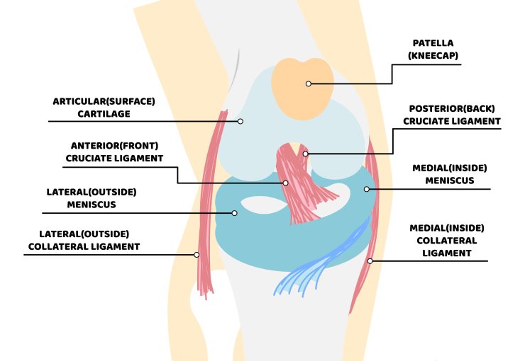
To accommodate extreme mechanical loads and complex multi-planar stresses, the knee joint is stabilized by four primary ligaments. Two collateral ligaments (MCL and LCL) provide external lateral and medial support, while the cruciate ligaments (ACL and PCL) cross within the joint center. The Anterior Cruciate Ligament (ACL) connects the femur (thigh bone) to the tibia (shin bone), acting as the primary anatomical restraint against anterior tibial translation and internal tibial rotation.
Surgical Management of ACL Tears
Anterior Cruciate Ligament (ACL) tear management is tailored to the patient’s athletic demands, age, occupation, joint laxity, and secondary lesions such as meniscal or chondral tears. In active individuals and athletes wishing to resume pivoting sports, anatomical surgical reconstruction is the gold standard.
Following acute ACL rupture, acute hemarthrosis and pain typically subside within 2–3 weeks. However, joint instability persists during rotational maneuvers, predisposing the knee to secondary meniscal tears and premature osteoarthritis if left unreconstructed.
Treatment follows an individualized clinical rule: evaluate the patient, not just the MRI. Concomitant meniscal repair or cartilage restoration is performed concurrently during arthroscopic reconstruction to maximize long-term joint health.
Clinical Risks of Chronic Instability
The knee is formed by the articulation of three major bones (femur, tibia, and patella), enabling smooth standing, walking, bending, and dynamic athletic movement while bearing substantial mechanical body weight.
Running obliquely within the center of the joint, the anterior cruciate ligament provides primary resistance against anterior tibial displacement and rotational pivot-shift instability.
Acute rupture presents with an audible “pop,” rapid hemarthrosis (joint swelling within hours), and inability to continue pivoting sports.
Without timely anatomic reconstruction, chronic ACL deficiency leads to recurrent giving-way episodes, progressive meniscal fraying, cartilage shearing, and premature post-traumatic osteoarthritis.
Diagnostic Evaluation & Clinical Pitfalls
In the acute post-injury phase, severe hemarthrosis and protective muscle spasm can hinder manual examination. High-resolution MRI confirms ACL disruption and details concomitant meniscal or chondral injuries.
Radiographs rule out avulsion fractures (such as a Segond fracture), while definitive diagnostic arthroscopy confirms ligament continuity and guide anatomical graft positioning.
Graft Choices in ACL Reconstruction
With the rise in recreational and competitive sports participation across all ages, individualized graft selection—matching the patient’s athletic profile—is fundamental to durable stability.
Graft Options: Hamstring tendon autografts provide exceptional tensile strength with minimal donor site morbidity, while Bone-Patellar Tendon-Bone (BPTB) and Quadriceps autografts provide robust osseous fixation.
Anatomic ACL reconstruction utilizes high-strength autografts—such as Hamstring tendon autografts, Bone-Patellar Tendon-Bone (BPTB), or Quadriceps tendon autografts. Prof. Dr. Yavuz Kocabey’s innovative Kocabey PressFit Technique achieves secure anatomical biological fixation without foreign screws, promoting rapid bone-to-bone osseointegration and faster functional rehabilitation.
For detailed information and appointment scheduling, please visit our Contact page.
Contact Us for Advanced ACL Treatment & Consultation.